Leja counting chamber 20 micron 2 chambers
In stock

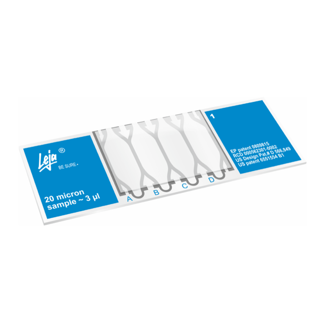
Leja counting chamber 20 micron 2 chambers
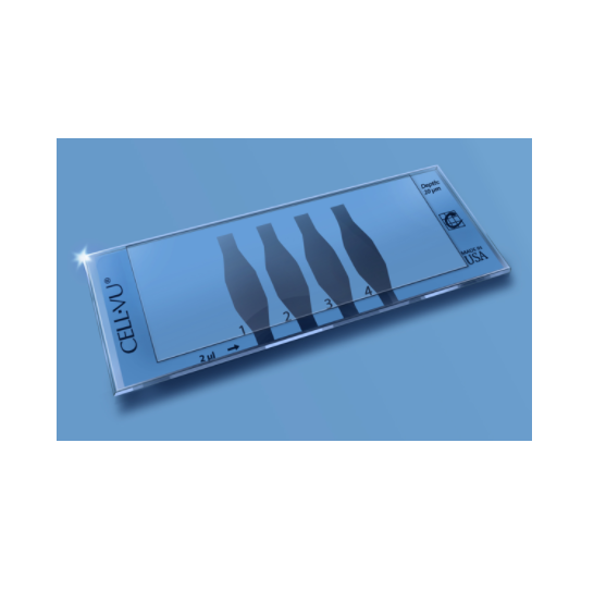
The Leja counting chamber has a thickness of only 10µm, meaning a tenth of a common haemocytometer, it is the flattest among all known counting chambers. Above all this counting chamber is used for sperm analysis. Spermatozoa move in one focal plane.
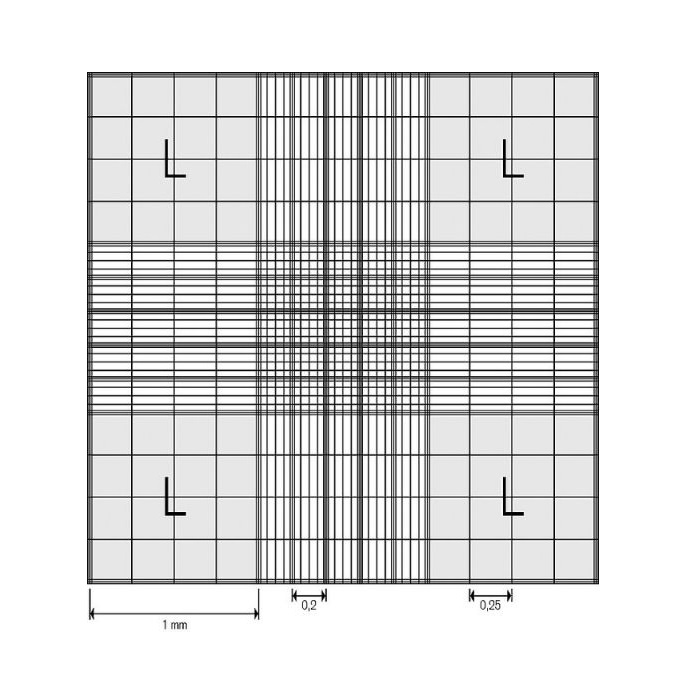
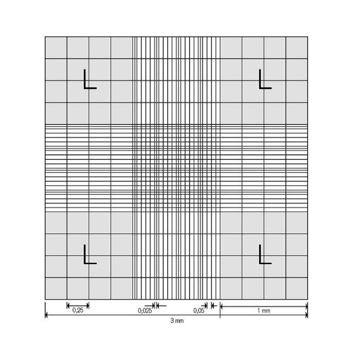
The counting chamber provides a grid with 10 x 10 squares. The size of one square is 100 µm x 100 µm.
Product facts and notices
- SpermCount:
The sample has to be liquefied and mixed homogeneously.
Avoid crystallization by leaving the sample stationary for a too long time.
Put a drop of the sample into the chamber and count the sperm heads as follows: 10 squares of the field are counted like blood cells in a haemocytometer. This number represents the concentration of spermatozoa in millions per millilitre.
When counting a sample of oligozoosperms all 100 squares have to be counted. Adding “00 000” to that number, you will get the concentration of the spermatozoa per millilitre.
- Determination of motility:
Immotile spermatozoa are to be counted in a defined number of squares. Afterwards the motile spermatozoa are counted and classified into four categories from a) “fast progressive” to d) “no motility”. This procedure is to be repeated in different areas of the grid to have the quality and motility to be calculated on base of this data in percent.
Features
- The Leja chamber is easy to use and you gain results fast and easy. The number of counted spermatozoa for each 10-field-grid is their concentration in millions/ml. For the calculation no conversion factors are needed.
- Optimal depth. The meagre depth of only 10 µm ist especially adapted to the size of the spermatozoa wich allows them to diffuse evenly in a monolayer. They can move about freely in a single focal plane without the picture to bedome indistinct.
- The grid is located in the cover glass. You don’t need a grid in your ocular.
- A dilution of the sample is not needed as the analysis is made with the original sample in its natural seminal fluid or in a concentrate.
- The analysis is made very fast meaning the patient does not have to wait too long for the results.
- The analysis is precise because the steps you usually have to undergo with a haemocytometer are omitted and you are always working under reproducible conditions. Uneven pressure on the cover glass does not lead to mistakes.
There are no specifications


There are no report
You May Also Like
BRAND™ Counting Chambers, Neubauer, Improved, BLAUBRAND®, Without Clips Double Ruling
$ On demand
On demand
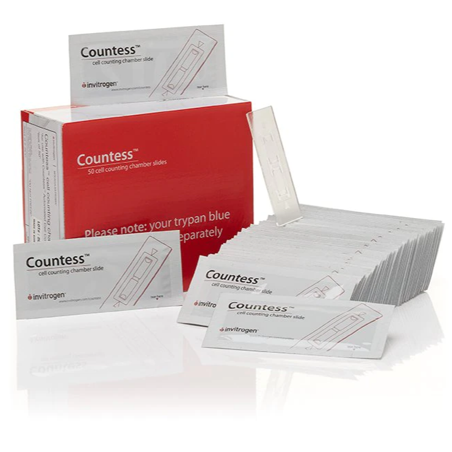
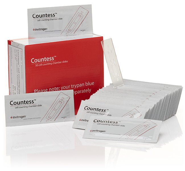
Invitrogen™ Countess™ Cell Counting Chamber Slides, 2,500 Slides
$ On demand
On demand
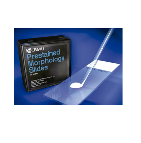
Hamilton CELL-VU® Fixed Coverslip Sperm Counting Chamber, No Grid, 2-chambers
$ On demand
On demand
BRAND™ Counting Chambers, Neubauer, BLAUBRAND®, Without Clips Double Ruling
$ On demand
On demand